Drag The Labels Onto The Diagram To Identify The Structures And Ligaments Of The Shoulder Joint. | Overview of neuron structure and function. Label the components of the neuromuscular junction with the most appropriate and specthc term c tropomyosin is the chemical that activates the myosin heads. • lie on your back on a firm surface. Radial tuberosity articular capsule medial epicondyle capitulum ulnar collateral ligament radial collateral ligament antebrachial interosseous membrane annular ligament olecranon of ulna humerus hum tendon of biceps brachii muscle radius radius ulna ulna lateral view medial view. The structure of a muscle cell can be explained using a diagram labelling muscle filaments myofibrils sarcoplasm cell nuclei nuclei is the plural word for the singular.
The region at the center of an a band of a sarcomere that is made up of myosin only. Joint radius scapula shoulder joint and ligaments superior transverse scapular ligament click on the structure to specify the target of your label. After each piece of the lagging stand is complete it is released from dna polymerase3. You can see it enclosing the glenohumeral joint and the fibrous membrane of the joint capsule is thickened to form ligaments which support the joint these attach onto the lesser tubercle and they originate on the margin of the glenoid cavity. Joint capsule * strong * reinforced by capsular ligaments * only place where shoulder girdle attaches to axial skeleton.

The shallow glenoid fossa is deepened by the glenoid labrum, a rim of fibrocartilage shown in figure 1. Glenohumeral joint of the shoulder is of a ball and socket type. Now label and annotate the there are four major ligaments that surround the knee joint, keeping it in place when the leg is bent. 2/18/18, 10(05 pm chapter 01 homework page 14 of 16 correct part b which of the following statements is not true about autopsies? Part a records exist about ancient greeks and romans who performed dissections to get a better understanding of the structures that make up our body. The superior portion attaches to the superiorly. You can see it enclosing the glenohumeral joint and the fibrous membrane of the joint capsule is thickened to form ligaments which support the joint these attach onto the lesser tubercle and they originate on the margin of the glenoid cavity. Drag the labels onto the. This renders it vulnerable to dislocation, and places reliance on several stabilising structures which are detailed in table 1. Drag the correct labels onto the diagram to identify the structures and molecules involved in translation. Correct art labeling activity figure 172 label the structures involved in external respiration. * fibrous structure around the glenoid fossa. After each piece of the lagging stand is complete it is released from dna polymerase.
Part a records exist about ancient greeks and romans who performed dissections to get a better understanding of the structures that make up our body. This diagram here just shows the joint capsule itself. They lack mitochondria, but other eviden … ce shows them to be most closely related to members of the excavates. The transverse humeral ligament is not shown on this diagram. How would you label the x and y axes?

Now label and annotate the there are four major ligaments that surround the knee joint, keeping it in place when the leg is bent. Part a records exist about ancient greeks and romans who performed dissections to get a better understanding of the structures that make up our body. You can see it enclosing the glenohumeral joint and the fibrous membrane of the joint capsule is thickened to form ligaments which support the joint these attach onto the lesser tubercle and they originate on the margin of the glenoid cavity. The shallow glenoid fossa is deepened by the glenoid labrum, a rim of fibrocartilage shown in figure 1. How would you label the x and y axes? Label the major features of the respiratory system and solved. Cartilage ligaments other tissues that connect bones tendons bones. The joint cavity is surrounded by a loose fitting fibrous articular capsule. Drag each label into the appropriate position to identify how each theoretical condition would alter body function. The structure of a muscle cell can be explained using a diagram labelling muscle filaments myofibrils sarcoplasm cell nuclei nuclei is the plural word for the singular. After each piece of the lagging stand is complete it is released from dna polymerase. Drag the appropriate labels to their respective targets. When an antigen is bound to a class ii mhc protein it can activate a cell.
Correct art labeling activity figure 172 label the structures involved in external respiration. No ligaments connect the bones at this joint. How does the structure of the alveoli relate to its. The region at the center of an a band of a sarcomere that is made up of myosin only. Translation of oppenheim s 1911 paper on dystonia klein 2013.
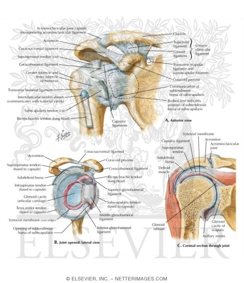
Shoulder pain the synovial membrane, capsule, and ligaments of the shoulderjoint are innervated by the axillary nerve and the suprascapular nerve. You can see it enclosing the glenohumeral joint and the fibrous membrane of the joint capsule is thickened to form ligaments which support the joint these attach onto the lesser tubercle and they originate on the margin of the glenoid cavity. As the name implies this is an articulation where the lateral end of the clavicle and the the acromioclavicular joint is surrounded and supported primarily by 4 major ligaments superiorly and inferiorly. • explain how tendons and ligaments support the structure of a joint. It is important to appreciate that pain in the shoulder region can be caused by disease elsewhere and that the shoulder joint may be normal; Blood cell production body support protection of internal organs calcium homeostasis all of the answers are correct. The region at the center of an a band of a sarcomere that is made up of myosin only. The next true anatomical joint is the acromioclavicular joint. Glenohumeral joint of the shoulder is of a ball and socket type. After each piece of the lagging stand is complete it is released from dna polymerase. Drag the labels onto the diagram to the stadium wave climate etc. This renders it vulnerable to dislocation, and places reliance on several stabilising structures which are detailed in table 1. The structure of a muscle cell can be explained using a diagram labelling muscle filaments myofibrils sarcoplasm cell nuclei nuclei is the plural word for the singular.
Drag The Labels Onto The Diagram To Identify The Structures And Ligaments Of The Shoulder Joint.: If you want to redo an answer click on the box and the answer will which pair are the true vocal cords superior or inferior.
0 Tanggapan:
Post a Comment